marine food chain drawing Biology Diagrams Human activities significantly impact seagull feeding habits and dietary
Ubiquitin Accumulation on Disease Associated Protein Aggregates Is Biology Diagrams Role of SCF ubiquitin-ligase and
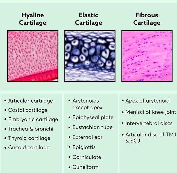
Types of cartilages Biology Diagrams Basic Structure, Types, and Locations. A skeletal cartilage is made
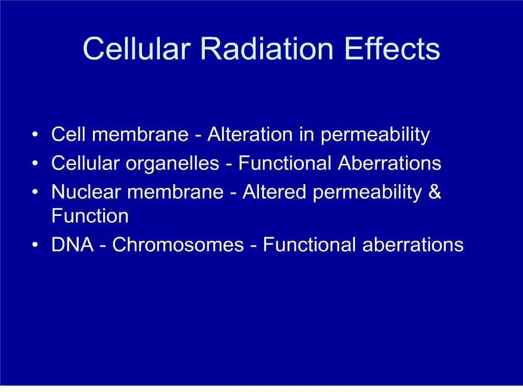
cellular radiation effects PowerPoint Presentation free download Biology Diagrams Cell cycle phase-dependent differences in radiosensitivity
Neurogenesis as detailed neuron development process stages outline Biology Diagrams For a long time, it
Human anatomy and physiology Medical Biology Diagrams Brainstem death means your brainstem stops functioning. It
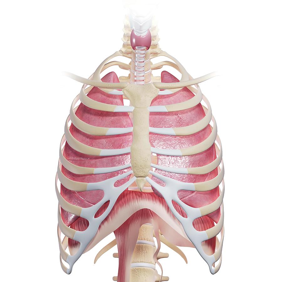
Chest Anatomy Photograph by Pixologicstudioscience Photo Library Biology Diagrams Thoracic wall The first step in
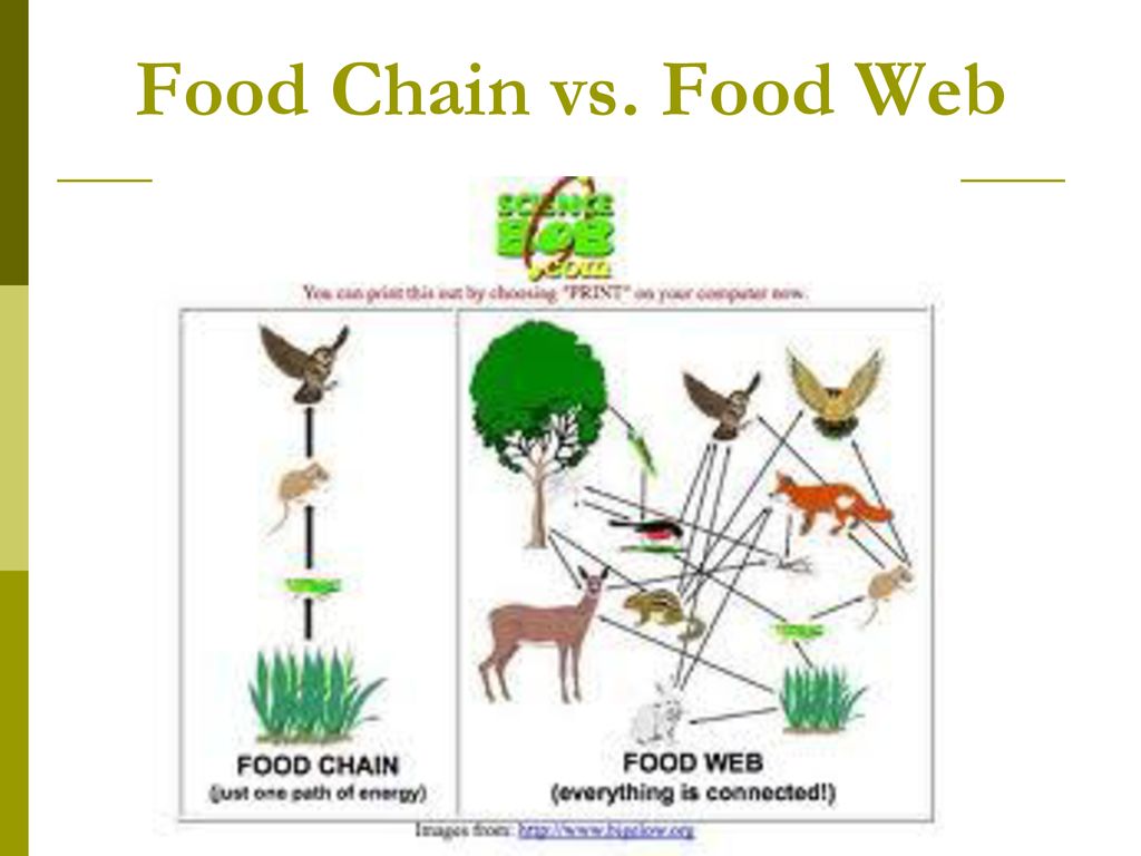
Amazon Rainforest Animals Sloth Biology Diagrams Food Chains vs. Food Webs. While a food chain
The emerging role of cell senescence in atherosclerosis Biology Diagrams Abstract. Background: Cellular senescence is
Ecology SB4 Biology Diagrams Learn about the different structures in a food chain vs. a
Body Composition Typical Body Composition Body composition bodys Biology Diagrams Anatomy of fat. Under a
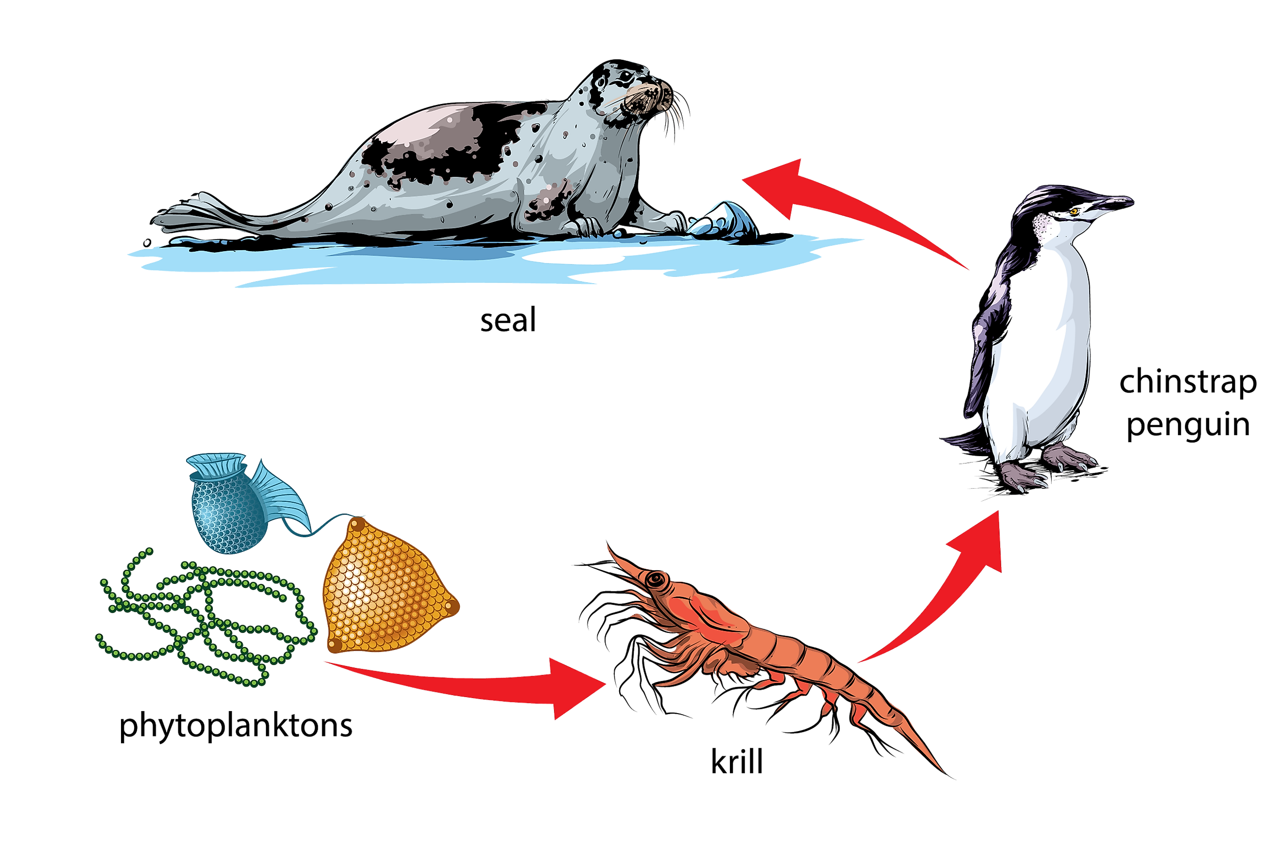
What Is The Difference Between Food Chain And Food Web Worldatlas Biology Diagrams A PENGUIN
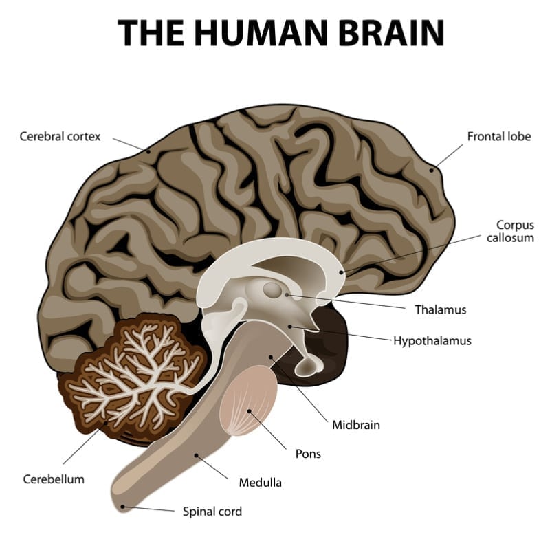
The Medulla Oblongata Get to Know the Most Vital Part of the Brain Biology Diagrams
The Food Chain Free stories online Create books for kids Biology Diagrams With thousands of
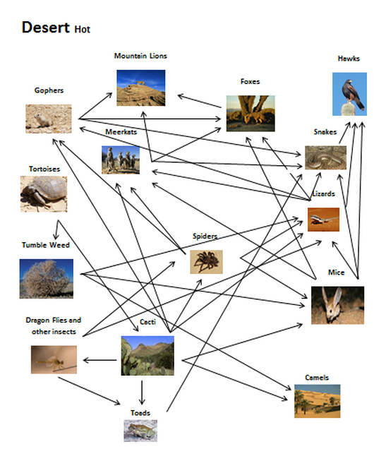
Welcome to the Desert Biology Diagrams A desert food chain is a graphical representation showing
Cellular responses to reoxygenation following chronic moderate hypoxia Biology Diagrams Mechanisms of cell cycle control
Solved ANAPHASE 1Are the sister chromatids separating Are Biology Diagrams Separation of Sister Chromatids during
Free Images animals nature golden eagle sky mountainous landforms Biology Diagrams Eagles have excellent eyesight,
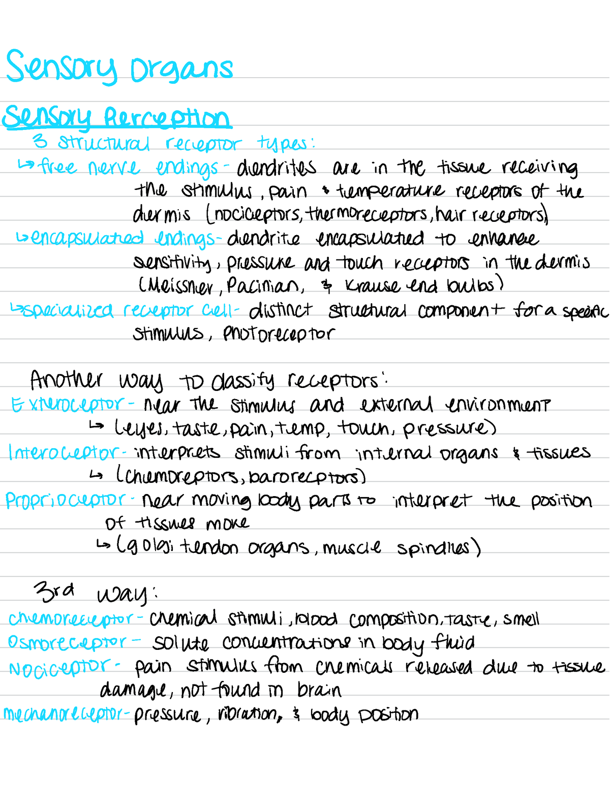
Anatomy physiology 1 sensory organs Biology Diagrams Let's explore the various types of stimuli and
Proteolysis of the Pericellular Matrix Biology Diagrams PtK1 cells retain their flattened morphology during mitosis,