Solved adrenal gland cortex medulla Figure 3 Adrenal gland Biology Diagrams Anatomical Structure. The adrenal glands consist of an outer connective tissue capsule, a cortex and a medulla.. Veins and lymphatics leave each gland via the hilum, but arteries and nerves enter the glands at numerous sites.. The outer cortex and inner medulla are the functional portions of the gland.
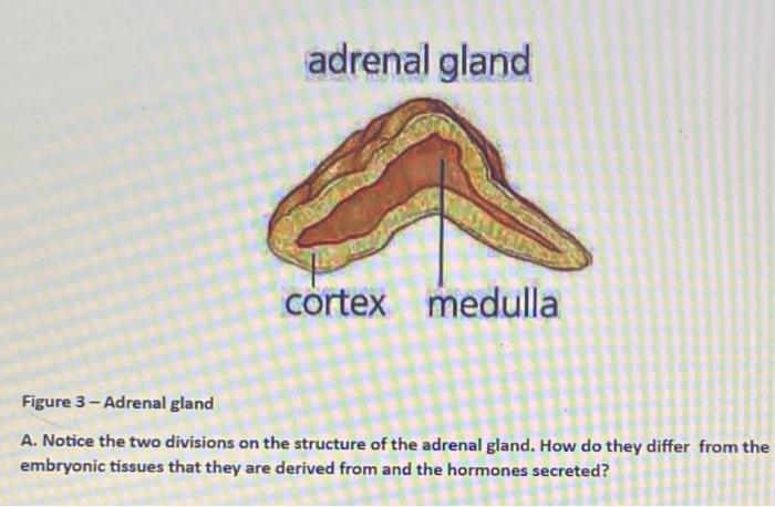
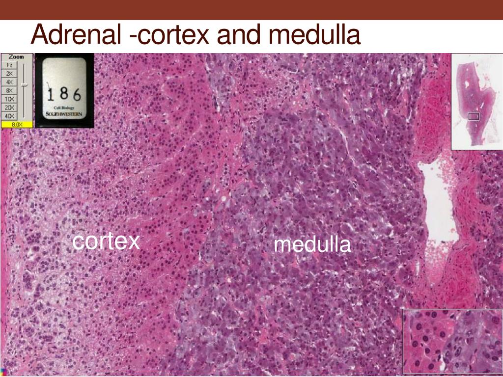
The adrenal cortex tends to be fattier and thus has a more yellow hue. The adrenal medulla is more of a reddish-brown color. A thick capsule consisting of connective tissue surrounds the entire adrenal gland. The adrenal cortex is much larger than the smaller medulla, accounting only for approximately 15% of the gland. These two major sections of the adrenal are called the adrenal medulla (inner layer), and the adrenal cortex (outer layer). The Adrenal Medulla: The adrenal medulla comes from the neural crest (i.e. embryological basis similar to your central and peripheral nervous systems). It contains homogenous sheets of cells organized into nests.
Anatomy, Abdomen and Pelvis: Adrenal Glands (Suprarenal Glands) Biology Diagrams
Anatomy . As mentioned, the adrenal glands are two small, roughly triangular glands that sit directly on top of the kidneys. The two major parts of the adrenal gland are the cortex and the medulla. The gland is surrounded by an adipose (fatty) capsule, which acts as a protective barrier.

Adrenal medulla. The adrenal medulla is embedded in the centre of the cortex of each adrenal gland. It is small, making up only about 10 percent of the total adrenal weight. The adrenal medulla is composed of chromaffin cells that are named for the granules within the cells that darken after exposure to chromium salts.

Adrenal Cortex: What It Is & Function Biology Diagrams
What is the anatomy of the adrenal cortex? You have two adrenal glands — one on top of each kidney. They contain the cortex, the largest part of the gland, and the medulla, the smaller inner part. Your adrenal gland is highly vascular, with as many as 50 arterial branches providing its blood supply. When does the adrenal cortex develop? Blood first flows through the adrenal cortex and then drains into the adrenal medulla. Adrenal hormones are released into the circulation via the left and right suprarenal veins. Figure 17.6.1 - Adrenal Glands: Both adrenal glands sit atop the kidneys and are composed of an outer cortex and an inner medulla, all surrounded by a connective
